Nucs AI's DeepPSMA and TrackPSMA Used by NIH in a Prospective Study of Biochemically Recurrent Prostate Cancer
The National Institutes of Health (NIH), through the National Cancer Institute (NCI), is using DeepPSMA and TrackPSMA in a prospective study of biochemically recurrent prostate cancer: a rising PSA after surgery or radiation that indicates residual or recurrent disease, in men with no metastases on conventional imaging.
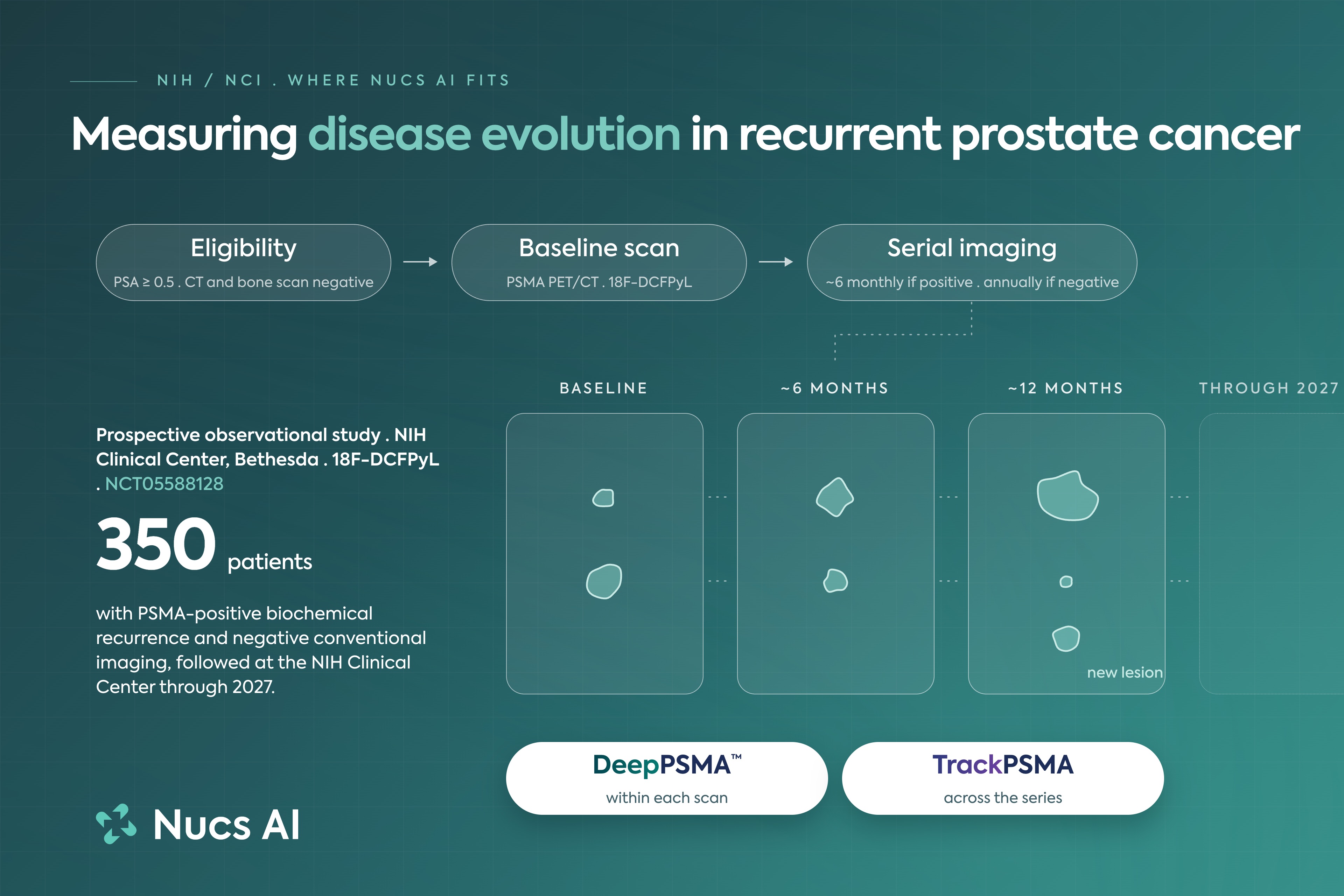
Measuring disease evolution in recurrent prostate cancer
The National Institutes of Health (NIH), through the National Cancer Institute (NCI), is using DeepPSMA and TrackPSMA in a prospective study of biochemically recurrent prostate cancer: a rising PSA after surgery or radiation that indicates residual or recurrent disease, in men with no metastases on conventional imaging.
The clinical problem
PSMA PET can find recurrent disease at PSA levels where CT and bone scan see nothing, by imaging a protein expressed on the surface of prostate cancer cells. Men who would once have been told they had no visible disease now have lesions on a scan.
The question is which of those men will progress, and which can be watched rather than treated. Only a minority develop metastatic disease. In the rest the disease stays indolent, often for the rest of their lives, and no imaging measurement reliably separates the two. So lesions on a scan are often met with therapy intended for metastatic disease, much of it given to men who would never have needed it, and androgen deprivation and radiation carry real cost. The smaller group who would genuinely benefit from treating early is not reliably identified either.
Nothing available answers it. Risk stratification still rests mainly on PSA level and PSA doubling time, both of which predate PSMA PET and were validated against conventional imaging. PSMA PET changes management for a majority of patients in prospective series, and guidelines note that whether those changes improve outcomes has not been established. The imaging criteria that do exist were built for advanced disease and for judging response to treatment, not for low-volume recurrence being observed rather than treated.
What the field lacks is an evidence base for watching. Thresholds that would make observation defensible, and a defined point at which observation should stop. Active surveillance works in low-grade localized disease because those thresholds exist. Nothing equivalent exists here.
Underneath all of it sits a more basic question: whether the trigger to stop watching should be how much disease is on the scan or how fast it changes between scans. With PSA, the rate of rise proved a stronger predictor of metastasis than the absolute level. Whether the same holds for imaging is unknown.
Answering that needs the same lesions measured the same way across years, which no dataset has done at scale, because until PSMA PET this disease was not visible.
The study
This is a prospective natural history study, conducted at the NIH Clinical Center by National Cancer Institute investigators, open since March 2023 and enrolling toward a target of up to 350 participants, each followed for up to five years. It is one of the few longitudinal PSMA PET datasets in this population.
The study team calls the population PSMA-positive biochemical recurrence, a term chosen to retain what is already known about the prognosis of biochemical recurrence. These men would not have had detectable disease, and would not have been eligible for metastatic disease trials, before PSMA PET existed. Most are observed off therapy, with short courses of treatment permitted while they remain on study.
Eligibility requires prior definitive treatment by surgery or radiation, PSA of at least 0.5 ng/mL, non-castrate testosterone above 100 ng/dL, and no metastatic soft tissue disease on CT or MRI and no bone lesions on bone scan. Participants with any findings, including equivocal ones, undergo repeat PET/CT with 18F-DCFPyL (piflufolastat F 18), approximately every six months and no more than twice a year. Those with negative findings are imaged annually. Conventional imaging is repeated annually and PSA tracked in parallel.
Study record: clinicaltrials.gov/study/NCT05588128
What DeepPSMA and TrackPSMA do
Hundreds of scans, years of follow-up and multiple readers mean hundreds of hours of manual outlining and extraction, and every inconsistency in how a lesion was measured becomes noise a real progression signal has to rise above. Nucs AI standardizes the measurement step: one automated method applied to every scan in a series.
DeepPSMA detects, stages, and quantifies a single scan. AI-driven tumor detection and segmentation at the level of individual image voxels, then lesion count, lesion volume, SUVmean, SUVmax and SUVtotal, reported at whole-body level, by compartment and for individual lesions. Compartments follow miTNM, the molecular imaging staging system defined in PROMISE.
TrackPSMA works on the series, connecting every lesion from one scan to the next. The same measures compared between timepoints at whole-body level, at TNM level across prostate, pelvic nodal, distant nodal, bone and visceral compartments, and for individual lesions. What it adds beyond comparison is correspondence: following the same lesion from one timepoint to the next, so a disease site has a trajectory.
Automated AI output is physician-reviewed before it is finalized.
The measurement layer
This measurement problem is not specific to prostate cancer. Any question that depends on comparing imaging over time meets it, and the same approach applies to other tracers and tumor types. That is the layer Nucs AI is building.
The open question is which men will progress, which have disease that never will, and how long a man can safely be watched. No single scan answers it. Years of consistent measurement might, and that is what this study is for.
Nucs AI supports research advancing molecular imaging and theranostics. Reach out at nucs.ai/contact